Cryobiopsy and Lung Cancer Diagnosis
- SABEST VIT 2021

- May 5, 2021
- 2 min read
- Srimathi L
Diagnostic histopathological studies help us form a better understanding of the nature of malignant growth in human cells and in determining the underlying disease. Such studies are crucial for the diagnosis and treatment of several forms of cancer. Cryobiopsy has proven to be an effective technique in retrieving sizeable samples for diagnosis of lung cancer, one of the most prevalent types in smokers globally.
Lung cancer is classified into small cell lung cancer (SCLC) and non small cell lung cancer (NSCLC). NSCLC is further divided into adenosarcoma, squamous cell carcinomas and large cell carcinomas. Prognosis for adenosarcoma is precise since it is one of the most common types of lung cancer. However, in order to diagnose the type of lung cancer correctly, there is a requirement for methods that do not lead to formation of crush artifacts (morphological alterations happening to the sample cell due to external factors) or iatrogenesis-diseases caused due to medical procedures like sample extraction and diagnosis. Hence, cryobiopsy plays a major role in treating pulmonary disease.
Bronchoscopic cryobiopsy is based on the Joule-Thomson phenomenon, which states that a real gas undergoes a decrease in temperature when there is a pressure drop. In simple words, a pressure- drop leads to the expansion of the gas and its subsequent cooling. The Bronchoscopic Lung Cryobiopsy (BLC) equipment is designed in accordance with this phenomenon. The equipment comprises of a console, a cryogen and a cryoprobe. The console helps monitor the pressure, the temperature at the tip of the probe and the time duration; the cryogen (usually carbon dioxide or nitrous oxide) which is responsible for cooling (−70 ◦C to −89 ◦C) and the cryoprobe which attaches to the tissue and collects the sample. This happens due to the rapid cooling of the area surrounding the tip of the probe, leading to the adhesion of the tissue around the probe. Later, the tissue is collected from the probe after multiple freeze-thaw cycles. Disposable cryoprobes have largely reduced the risk of cross-contamination.
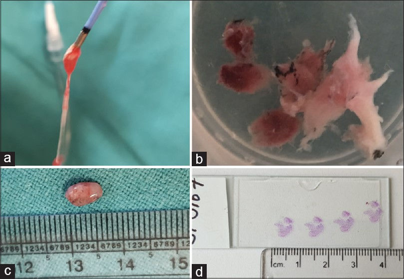
Image credits- https://lungindia.com/article.asp?issn=0970-2113;year=2021;volume=38;issue=2;spage=109;epage=116;aulast=Goel
From image- From left to right: Tissue sample attached to the probe, sample isolated from probe for diagnosis, size of the sample obtained, stained samples ready for observation under the microscope.
Cryobiopsy helps obtain a better sample size that can be well preserved for peripheral and endobronchial tumours. Research has shown that the diagnostic yield of cryobiopsy as opposed to traditional forceps biopsy is significantly higher. The greater size and volume of the collected sample coupled with absence of high amounts of crush artifacts increase the chances of precise diagnosis and treatment of lung cancer in its early stages. The technique, however, has one drawback. Since the cryoprobe is stiff, it takes some time to make adjustments so that it reaches the target tumour and attaches to it, thus prolonging the duration of the procedure.
In conclusion, cryobiopsy is an effective technique that can be used in the diagnosis of lung cancer. If combined with other methods like fluorosopy, it can increase the diagnostic yield of samples collected from suspected endobronchial cancer/peripheral cancer patients, leading to precision treatment that targets only the diseased cells. Cryobiopsy, thus, can play a potentially life-saving role in future lung cancer diagnostics.
For more, visit: https://pubmed.ncbi.nlm.nih.gov/33921579/


תגובות